 Created by renowned Harvard health care professionals.
Anatomy of the Carpal Tunnel & Surrounding Soft Tissue
We rarely think about how much is happening inside our hands, or how seamlessly everything works together—until there is a problem. While bones give the hand its structure, it’s the soft tissue—tendons, ligaments, muscles, and lymphatic channels—that allows it to function pain-free. Dive into a structure-by-structure breakdown of the carpal tunnel to understand how these tightly packed components work together, and why a lack of anatomical slack makes the hand so susceptible to Carpal Tunnel Syndrome.

Anatomy of the Carpal Tunnel and Soft Tissue That Allows the Hand to Work
Every time you type, grip, pinch, or bend a finger, you rely on a remarkably compact piece of anatomy at the base of your palm: the carpal tunnel.
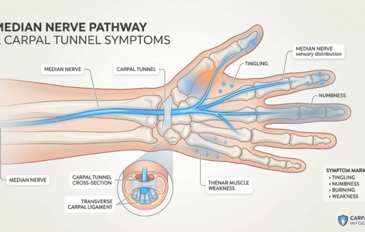
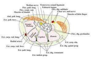
This small, rigid passageway contains more than 20 distinct soft tissue structures working in incredibly close quarters. All of it—the median nerve, nine flexor tendons, lubricating sheaths, stabilizing ligaments, flanking muscles, and a network of blood vessels and lymphatic channels—shares a space that cannot expand. For your hand to work, these structures must glide together in perfect, friction-free sync.
It is precisely because this space is fixed that Carpal Tunnel Syndrome (CTS) happens at all. When any of these tightly packed structures swell, the space runs out. Since the median nerve is the softest, most pressure-sensitive structure in the tunnel, it gets squeezed first. That pressure is what produces the trademark numbness, tingling, and weakness of CTS.
What We Mean by "Soft Tissue"
Throughout this website, you will often see the term “soft tissue.” This refers to every structure in the hand and wrist other than bone: tendons, ligaments, nerves, muscles, blood vessels, lymphatic channels, and the connective tissue supporting them.
Bones themselves rarely cause CTS. It is the soft tissue that becomes inflamed, swells, and creates nerve pressure. The lymphatic channels are especially critical here; they act as the body’s drainage system for excess fluid and inflammatory byproducts. When they become sluggish or overwhelmed, fluid has nowhere to go but into the surrounding tissue, increasing the pressure inside the tunnel.
A Crowded Space: The Anatomy Breakdown
To understand why the wrist is so vulnerable to injury, it helps to look at exactly how many moving parts are packed into and around the carpal tunnel.
Transverse Carpal Ligament: A tough fibrous band stretching across the wrist.
Converts the wrist bones into a closed, rigid tunnel. If soft tissues swell, this unyielding roof forces the pressure downward onto the nerve.
Carpal Bones & Ligaments: A shallow arch of small wrist bones held together by short intercarpal ligaments.
Role in CTS: Forms the hard base of the tunnel. Without these ligaments, the arch would splay, but because they hold the bones tight, the tunnel’s volume remains strictly fixed.
The Median Nerve & 9 Flexor Tendons: The cords that bend your fingers and thumb, plus the nerve that provides feeling to them.
Role in CTS: These 10 structures are the tunnel’s primary occupants. Repetitive strain can cause the tendons to inflame (tenosynovitis), directly crowding and crushing the delicate median nerve.
Synovial Sheaths & Connective Tissue: Slippery tubes wrapped around the tendons (ulnar and radial bursae) and loose tissue filling the gaps.
Role in CTS: Allows tendons to glide without grinding. If these sheaths swell with fluid or inflammation, they rapidly consume what little free space is left in the tunnel.
Thenar & Hypothenar Muscles:The muscle groups at the base of the thumb and pinky.
Lumbrical Muscles: Deep hand muscles.
Role in CTS: While mostly outside the tunnel, certain wrist positions can cause these muscle bellies to bulge inward, temporarily increasing pressure on the median nerve.
Vascular & Lymphatic Channels: Arteries, veins, and the vasa nervorum (tiny vessels feeding the nerve), plus the lymphatic drainage network.
Role in CTS: If lymphatic drainage slows, fluid pools in the wrist. Additionally, prolonged pressure inside the tunnel can cut off blood flow to the nerve itself, causing ischemic injury and severe pain.
Guyon’s Canal & Palmar Nerves: A separate nerve/artery pathway next to the tunnel.
Role in CTS: Explains why CTS usually spares the pinky finger (fed by Guyon’s canal) and the palm’s surface skin (fed by a nerve branch traveling over the tunnel roof).
The Vulnerability of a Perfect Machine
The hand and wrist contain 27 bones and dozens of moving parts. It is easy to take for granted, but it is the soft tissue that cushions and guides every movement so the hand can function pain-free.
This tightly packed, fixed-volume design allows for incredible precision, but leaves zero margin for error. There is no anatomical slack—no extra room for a thickened ligament, a swollen tendon sheath, or an accumulation of fluid. Whatever inflammation the soft tissue receives, whether from repetitive strain, trauma, or a systemic condition, has nowhere to go but toward the nerve itself.
The hand can only heal when that trapped inflammation is addressed and the body’s natural drainage systems can clear the excess fluid from this incredibly crowded space.